When the blood becomes “saturated” with metabolic acids beyond the buffer system’s ability to eliminate, along with chronic inflammation, excess acids get deposited or stored in your fat cells, tissues, joints, and organs (like kidney, gallbladder, liver), arterial walls, and blood vessels, etc. In order to temporarily neutralize blood, interstitial fluid, and tissue acid your body will combine alkaline minerals like sodium, calcium, and sometimes magnesium with these acids to form crystal salts.
High acid producing diets and stress deplete available alkaline minerals fairly rapidly, FORCING the body to pull what it needs from emergency stores like the bones and muscles. For example, low dietary and supplemental magnesium in conjunction with stress, substantially increases the risk of crystal calcification over time, especially when calcium/magnesium levels are grossly disproportionate, which is VERY common.
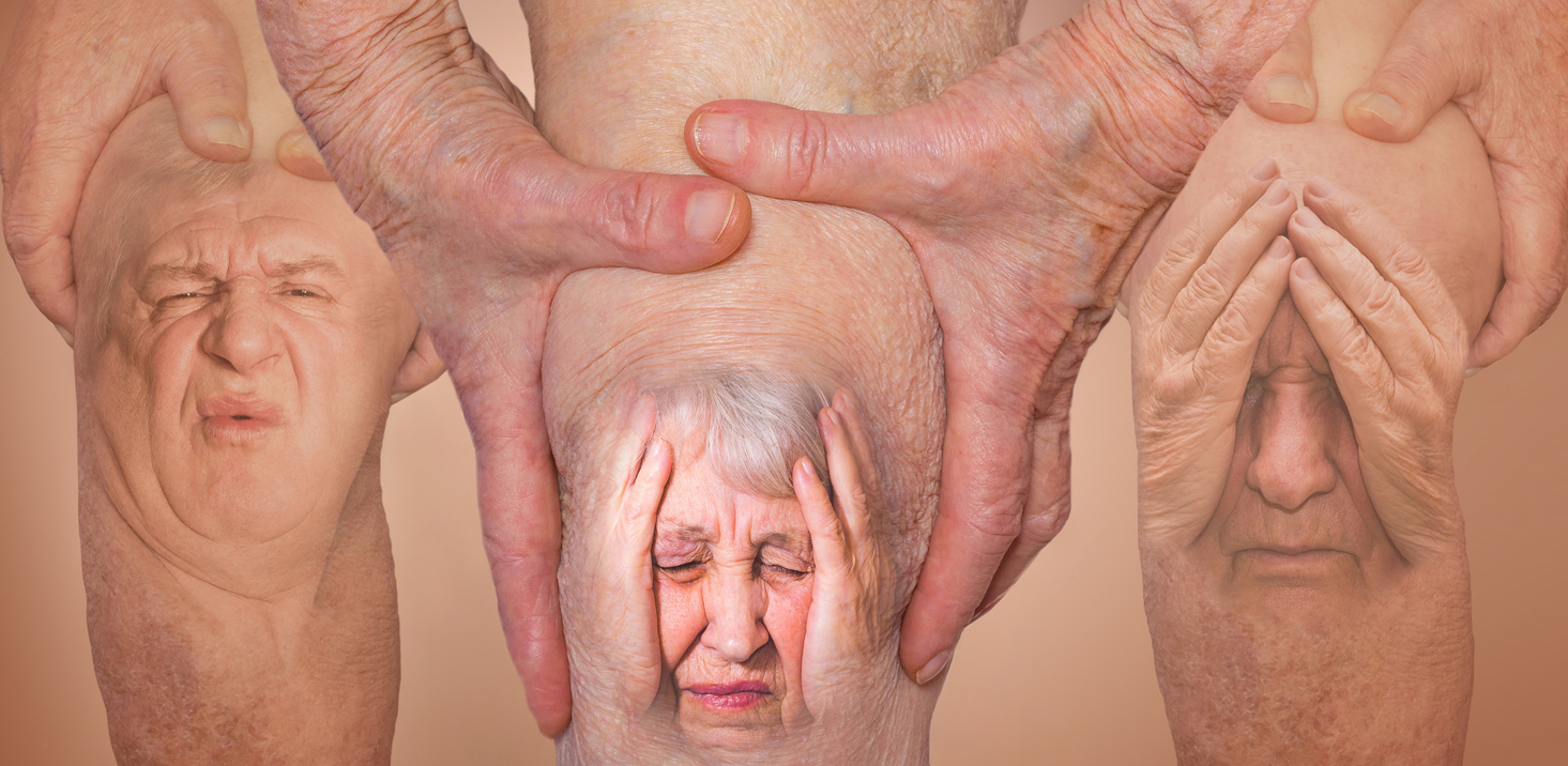
When the body builds-up with these acids and calcified salts, you will likely experience various symptoms and issues, such as fatigue, weight gain, nerve disorders, joint problems, heart and liver problems, kidney disorders, prostate issues, breast problems, you name it. Previously injured, overworked joints, and arthritic and damaged tissues are more vulnerable to crystallization destruction, ESPECIALLY in a chronic inflammatory acidic environment.
Conventional medicine once again provides NO answers, and claims that these conditions are not curable, which is far from true in most cases. For joint issues, many conventional doctors recommend pain killers to temporarily relieve pain and inflammation at the expense of your liver, kidneys, intestinal health, and other organs, with no real solution, only symptom management. This is a BAND AID approach, which can further destroy the joints. Then they offer surgery as a last resort when immobility sets in. Often times, these surgeries do not provide any long-lasting relief, and since the internal environment has not changed, it’s just a matter of time before the next joint or organ may suffer irreversible damage. Even diagnostically, conventional medicine provides confusing, conflicting, and incomplete information regarding the epidemiology, and is ESPECIALLY lacking in the etiology behind acid saturation and crystal deposition.
Instituting an anti-inflammatory pH balanced diet and lifestyle is the only way to assure the elimination and further deposition of damaging liquid acids and acid induced crystallization. When blood acid levels are low enough, some of the acid crystals can precipitate back into the blood for elimination. However, without instituting dietary and lifestyle changes the overly acidic environment will continue to present a danger to your health. When regularly consuming lemon water with bicarb and taking a few scoops of highly alkaline greens mix, the blood will be capable of accepting some of the acids from the tissues, giving you pain relief as inflammatory acids migrate out of the tissues into the blood for elimination!
ACID CRYSTALS ARE FAR MORE COMMON THAN THEY ARE DETECTABLE
Unbeknownst to conventional medical wisdom, studies have found that besides gouty arthritis and kidney stones, urate and other crystals are showing up in other forms of arthritis, as well as organs like the heart and prostate! So maybe serum hyperuricemia and the resultant interstitial fluid hyperacidity should be taken a little more seriously? After all, these urate crystals would not appear without having elevated uric and other acid levels in the first place.
Virginia Byers Kraus, MD, PhD, professor of medicine at Duke University in the Division of Rheumatology and Immunology and senior author of the following study stated, “This research is a step towards identifying uric acid as a risk factor for osteoarthritis.” The researchers looked at 159 people, who had knee osteoarthritis but no history of gout (urate crystal deposits). “The researchers found the severity of osteoarthritis in their knees to be strongly correlated with the amount of uric acid in their knees. The results, which are published online in the Proceedings of the National Academy of Sciences, show, despite the lack of gout history, above normal uric acid levels in 39 percent of the study population, and evidence of the form of inflammation in the joints that is typically triggered by uric acid crystals.” That percentage would be much higher if they tested the interstitial fluids as well as the blood.
“In a non-gout population, this provides some of the very FIRST evidence that uric acid level is a potential cause of inflammatory events and joint degeneration in osteoarthritis,” Kraus said. (1)
In a 2014 observational study ‘Prevalence of birefringent crystals in cardiac and prostatic tissues’, published in the BMJ medical journal authors assessed the prevalence of urate crystals in residual tissue samples in coronary arteries from 55 explanted hearts, 75 aortic valves collected from valve replacement surgeries, and 40 frozen, unfixed prostate specimens.
By definition, hyperuricemia interstitial fluids are supersaturated, and urate supersaturation leads to precipitation of urate crystals. In joints, the crystals often drive the inflammatory disease recognized as gouty arthritis. However, hyperuricemia is not limited to plasma and the joints. All other interstitial fluids (excepting cerebrospinal fluid and sweat) will be supersaturated as well. Consequently, urate crystals will occur in non-articular (other than joint) tissues and may well drive crystal-induced inflammation.
The authors claim, “This was the first study to systematically evaluate for the prevalence of urate crystals within tissues.” It was also the first study to show a significant percentage of “outside the joint” more problematic negatively birefringent (asymmetrical) crystals.
Researchers concluded:
a remarkable percentage of coronary arteries and prostate specimens contained birefringent crystals. Crystal presence is an obvious prerequisite for possible crystal induced-inflammation in these tissues, just as similar crystals elicit a gouty inflammatory cascade in synovial joints. Further studies are necessary to determine whether urate crystals may play this role in these tissues and, if so, to establish whether urate-lowering therapy may be beneficial in prostatitis and coronary disease.(2) http://bmjopen.bmj.com/content/4/7/e005308.full -crystal pics available on this site.
TYPES OF CRYSTALS AND DIAGNOSTIC METHOD
The crystals most commonly recognized in synovial fluid analysis are the MSU (mono sodium urate) crystals most frequently involved in gout, CPPD (calcium pyrophosphate dehydrate) crystals involved in pseudo gout, and BCP (basic calcium phosphate) in conjunction with (HA) Hydroxyapatite (bone), a BCP compound. All of these crystal types have been found to form together at the same time in the same area of the body, annunciating the underlying acid saturation issue. For example, (CPPD) deposition disease can CO-EXIST with gouty arthritis (MSU crystals), osteoarthritis, and bacterial joint infections. In fact, it is estimated that 20% of patients with CPPD also have hyperuricemia and 25% of these patients will develop gout. Remember, that is a very low percentage with hyperuricemia only recognizing blood levels, not tissues and interstitial fluids.
Compensated polarized light microscopy (CPLM) uses light microscopy to identify type of crystals by their birefringent (asymmetrical) properties, based on certain color patterns when rotated under a microscope. This method can be used to aide in the diagnosis of arthritis induced by MSU and CPPD crystals, as both of these crystal types show some birefringence and also have noticeably different appearances. Gout crystals (MSU) are needle shaped, but pseudo gout crystals (CPPD) are mainly rhomboid shaped. All MSU crystals are highly asymmetrical and can be easily recognized by use of CPLM. However, CPPD crystals are only slightly birefringent which is why CPPD crystals are only detected 20% of the time. With somewhat limited resolution of CPLM, besides CPPD even smaller crystals of MSU can be hard to pick up unless there are a lot of them, making early detection near impossible.
In contrast to MSU and CPPD, BCP crystals mostly present themselves as not birefringent, often misidentified and cannot be seen through polarized light microscopy unless crystals are abundant. Therefore, although not identified, very small BCP crystals could easily be the culprit even when a crystal-related arthritis patient’s synovial fluid is void of asymmetrical rods or needles.
MSU CRYSTALS AND GOUT
Gout is sometimes referred to as CRYSTAL ARTHRITIS, distinguished by the supersaturation, precipitation, and deposition of MSU crystals in joints and tissues, producing inflammation, resulting in tissue, joint, and bone damage. MSU crystals in particular, are far less soluble than uric acid by itself and deemed the primary culprit of gout and other degenerative joint conditions.
Besides the irritation from the sharp crystals themselves, when MSU crystallizes in the joints for instance, the immune system produces various white blood cells to initiate an attack on the invading foreign crystals. Correspondently, inflammation develops, producing swelling, shiny red or purple extremely tender skin, along with fierce and unbearable pain, often accompanied by weakness, lethargy, increased heart rate, high blood pressure, fever, and chills. These incidences can happen suddenly without warning!
Gout and other forms of arthritis take place mostly in the joints- toes, ankles, knees, arms, elbows, hands, wrists, bursas and sometimes shoulders, necks, hips and spine, as well as the ligaments and tendons that surround these joints. These crystals can invade MULTIPLE joints or other areas at the same time. Some individuals with gout may only experience 1-2 attacks per year or less, depending on corrective measures taken. For others, however, the gout can be chronic in nature, recurring regularly with consistent chronic inflammation, causing significant degradation and deformities similar to rheumatoid arthritis. Gout attacks often occur between 12 and 8 a.m. presumably due to lower body temperature, relative nocturnal dehydration, or the nocturnal dip in cortisol levels and more frequent in spring than other seasons. With advanced gout, tophi urate crystal deposits can be found under the skin, joints, bursa, and ears leading to the destruction of bone and cartilage. They can even be found in the heart and other organs.
TEMPORARY GOUT REMISSION – TWO DIFFERENT THEORIES
THEORY #1-Interestingly enough, these crystals can go into hiding. After a gout attack, MSU crystals can be coated by certain proteins called apolipoproteins. The crystals then become unrecognizable to the immune system and become embedded inside the joints and tissues. Cancer cells, viruses, and bacteria do something similar, but more as a protective mechanism for survival purposes. When the crystals are camouflaged the gout then goes to remission. When conditions are right, these crystals shed their coatings and once again are recognized by the immune system as foreign, so white blood cells initiate an ATTACK once again reigniting the inflammation.
The gouty arthritis and the resultant degradation continues until the crystals are again coated and this VICIOUS cycle continues. When these old crystals shed their protein coating gout attacks occur whether the blood is hyperuricemic or not. Yes, new gout can be caused by the precipitation of new accumulating acids and crystals, but attacks are more likely triggered by the shedding of old crystals. This is why some people who temporarily go on a low purine diets are still at increased risk for recurring attacks. The following study backs this theory.
As per a 1993 PubMed study, ‘Changes in the proteins coating monosodium urate crystals during active and subsiding inflammation’, researchers performed an in vivo study investigating changes in the proteins that coat monosodium urate (MSU) crystals in human synovial fluid samples and rat air pouch fluid samples obtained sequentially during periods of active and resolving inflammation. These apolipoproteins formed on crystals as the inflammation subsided concurrently. In conclusion, the researchers stated:
This study provides the first in vivo evidence supporting the notion derived from previous in vitro studies that proteins coating MSU crystals change as inflammation evolves. IgG immune antibodies coating MSU crystals may enhance the inflammation. As the inflammation subsides, apolipoproteins could displace the IgG antibodies by competitively coating sites on crystals and could contribute in part to the resolution of the acute gouty arthritis.
This means the gout goes into remission as the proteins block immune activity. (3)
Even without treatment, initial attacks can subside within a few days to several weeks as the crystals become coated by these proteins, thus hiding from the immune system. Since conventional doctors struggle to eliminate the old crystals and prevent recurring attacks, they consider gout is to be incurable, believing it can only go into remission like most other diseases, which we know is FAR from true!
THEORY #2-Another well backed theory relating to chronic tophaceous gout (an advanced form of gout), involves additional more densely packed MSU crystals along with dead cell debris and white neutrophil (immune) blood cells surfacing years after the first diagnosis of gout. Along with the resultant inflammation, increased free radical activity, and acidity from these crystals comes a great deal of dead cell debris. As these cells die, DNA is extruded from the cells in large quantities, spilling purine rich nucleic acids, further increasing urate levels. When additional acids and cell debris come in contact with the MSU crystals, along with impressive amounts of neutrophil cells, it tightly packs the MSU crystals together and forms “a trap”. The neutrophil white blood cell consumption of MSU crystals, and the resultant DNA accumulation from dead cells involves a process called NETosis, or Neutrophil Extracellular Trap formation. The more neutrophils recruited to the sites of MSU deposits, the denser clusters of MSU crystals and formation of NET’s. This process is found to actually subdue inflammation through the degradation of pro-inflammatory cytokine immune cells. The following study confirms this scenario. (4)
In a 2014 Pub Med study, “Aggregated neutrophil extracellular traps limit inflammation by degrading cytokines and chemokines” authors explore this process. They found that, “Inflammation resolves spontaneously within a few days, although MSU crystals can still be detected in the synovial fluid and affected tissues. Here we report that neutrophils recruited to sites of inflammation undergo oxidative burst and form neutrophil extracellular traps (NETs). Under high neutrophil densities, these NETs aggregate and degrade cytokines and chemokines via serine proteases.“(5)
ALL references found in ACID CRYSTAL SALTS AND NEGATIVE IMPACT ON THE BODY section.